
Mastering microscopy techniques is no longer just a bonus—it’s a necessity. Whether you’re visualizing subcellular structures, tracking dynamic processes in live cells, or screening drug candidates at high throughput, the microscope you choose and the skills you develop can make or break your experiments.
Today’s modern microscopy tools are designed not only to push the boundaries of resolution and sensitivity but also to accelerate learning curves, streamline data acquisition, and democratize access to advanced imaging. Here’s how you can turbocharge your biotech skill set by leveraging these cutting-edge technologies.
1. From Widefield to Fluorescence: Building a Strong Foundation
At the heart of many biotech labs, the fluorescence microscope remains the workhorse for studying proteins, nucleic acids, and small molecules tagged with fluorescent probes. Fluorescence microscopy combines optical filters and light sources to excite specific fluorophores and collect emitted light against a dark background, yielding high contrast and specificity. Modern systems often include LED light engines, motorized filter wheels, and sensitive cameras (e.g., sCMOS) that dramatically reduce phototoxicity and bleaching, making live-cell imaging more accessible than ever.
Tip: Start by mastering fluorophore selection—understand excitation/emission spectra to avoid bleed-through, and learn basic filter set configurations. A clear grasp of these fundamentals will pay dividends when you advance to more complex systems.
You might wnat to learn more and find out more about fluorescence, You can find a very digestible guide just by searching online.
2. Confocal and Spinning Disk Systems: Stepping Into 3D
Once you’re comfortable with 2D widefield imaging, the next leap is into confocal microscopy. By introducing a spatial pinhole to reject out-of-focus light, confocal systems generate optical sections that can be reconstructed into crisp three-dimensional images. Spinning disk confocals accelerate this process by parallelizing pinholes on a rotating disk, enabling high-speed volumetric imaging of living specimens with minimal photodamage.
- Why it matters: 3D imaging is crucial for understanding tissue architecture, organoid development, or cellular interactions in real time. Investing time to learn confocal controls—such as pinhole size, Z-step calibration, and laser power modulation—will vastly expand your experimental repertoire.
- Further reading: For a deep dive into confocal principles and applications in cell biology, see the Nature Reviews Methods primer on modern fluorescence imaging techniques.
3. Super-Resolution Microscopy: Pushing Beyond the Diffraction Limit
Traditional light microscopy is limited to ~200 nm resolution by the diffraction of light, but super-resolution techniques such as STED, PALM, and STORM circumvent this barrier. While the instrumentation and image reconstruction pipelines are more complex, many commercial systems now offer turnkey modes that guide users through acquisition parameters and basic analysis workflows. Learning how to prepare samples (e.g., selecting photostable dyes), optimize labeling density, and process large datasets will make you a valuable asset in any imaging-driven project.
- Getting started: Look for workshops or online tutorials offered by microscopy core facilities or instrument vendors. Hands-on experience under the guidance of an expert can demystify calibration routines, alignment procedures, and post-processing in software like FIJI/ImageJ.
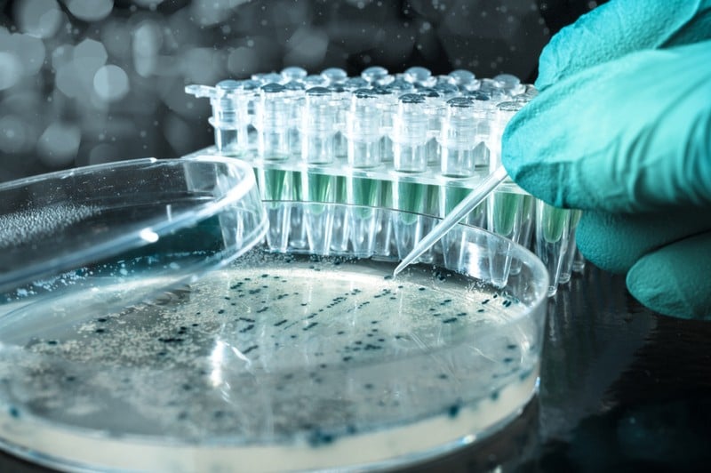
4. Automation and High-Content Screening: Scaling Up
As biotech shifts toward larger datasets, the ability to automate imaging tasks—like multi-well plate screening—becomes indispensable. High-content screening (HCS) platforms integrate plate handlers, robotic stages, autofocus routines, and analysis pipelines to collect and quantify phenotypic data across thousands of samples. Even if your lab doesn’t have a dedicated HCS system, many modern microscope software packages include scripting interfaces (Python, MATLAB) and application programming interfaces (APIs) that allow you to automate repetitive workflows, from time-lapse imaging to image-based cell counting.
- Skill to acquire: Basic proficiency in a scripting language (e.g., Python) can empower you to write custom macros for autofocus, z-stack acquisition, and batch analysis—transforming you from a passive user into a power-user who can tailor the instrument to your unique experimental needs.

5. Virtual and Augmented Reality: The Next Frontier in Training
Emerging educational tools are incorporating virtual reality (VR) and augmented reality (AR) to teach microscopy concepts in immersive environments. Instead of reading about optical paths on paper, you can don a VR headset and “walk through” the lens elements, light sources, and detectors in three dimensions. These platforms often include simulated hands-on modules where you can practice focusing, adjusting illumination, and troubleshooting common issues, risk-free, before you ever touch the real instrument.
- Why embrace it: VR and AR training modules can shorten onboarding time for new students and reduce wear-and-tear on precious optics by minimizing mistakes during initial familiarization.
6. Cultivating a Continuous-Learning Mindset
Finally, no amount of advanced instrumentation can replace the value of a curious, analytical mindset. Engage with online communities (e.g., Microscopy Listservs, Image.sc Forum), attend workshops at conferences (e.g., BISC, EMBO Imaging), and seek out interdisciplinary collaborations with physicists or engineers developing next-gen optical systems. By building a network of peers and mentors, you’ll stay abreast of emerging trends, such as adaptive optics, light-sheet microscopy, and AI-driven image analysis, and be ready to integrate them into your projects.
Conclusion
Accelerating your biotech skills using modern microscopy tools is an empowering experience that requires theoretical knowledge, hands-on practice, and collaborative problem-solving. From fluorescence basics to super-resolution imaging and automated screening systems, each step broadens your capacity to ask more advanced biological questions and answer them successfully. So embrace technology, form learning communities around it all, and keep in mind that the sharpest images often result from sharpest minds!